Peripheral angiography is a specialized diagnostic procedure used to visualize the blood vessels outside of the heart and brain, primarily focusing on the arteries that supply blood to the legs, arms, and other peripheral regions of the body. This imaging technique is crucial in diagnosing and assessing conditions such as peripheral artery disease (PAD), which can lead to serious complications if left untreated.
Peripheral angiography, also known as peripheral arterial angiography, involves the use of contrast dye and X-ray imaging to create detailed images of the blood vessels. The procedure allows doctors to see blockages, narrowing, or other abnormalities in the peripheral arteries, helping to determine the best course of treatment.
Peripheral angiography is typically recommended for patients who exhibit symptoms of peripheral artery disease or other vascular conditions, including:
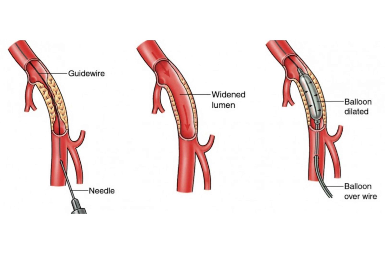
The procedure is usually performed in a hospital or specialized outpatient facility and involves the following steps:
The entire procedure typically takes about one to two hours, and patients are usually monitored for a few hours afterwards to ensure there are no complications.
Peripheral angiography offers several important benefits:
After the procedure, patients are usually advised to rest for a few hours to allow the incision site to heal. Drinking plenty of fluids can help flush the contrast dye from the body. Patients should follow their doctor’s instructions regarding activity restrictions and wound care.
Peripheral angiography is a valuable diagnostic tool in the evaluation and management of peripheral artery disease and other vascular conditions. By providing detailed images of the peripheral arteries, it allows for accurate diagnosis and effective treatment planning. If you are experiencing symptoms of PAD or have been recommended for a peripheral angiography, consult with your healthcare provider to understand the benefits and risks specific to your condition.
Peripheral angiography is a diagnostic imaging procedure used to visualize the blood vessels outside the heart and brain, particularly the arteries in the legs, arms, and other peripheral regions. It involves the use of contrast dye and X-ray imaging to detect blockages, narrowing, or other abnormalities in the blood vessels.
Peripheral angiography is typically recommended if you have symptoms of peripheral artery disease (PAD), such as leg pain or cramping while walking (claudication), non-healing wounds on your legs or feet, reduced or absent pulses in your limbs, or other signs of poor blood circulation. It helps in diagnosing the cause and extent of vascular issues and planning appropriate treatment.
Preparation for peripheral angiography may include:
During the procedure:
The procedure usually takes 1-2 hours.
You may feel a slight pinch or pressure when the local anaesthesia is administered and the catheter is inserted, but the procedure itself is generally not painful. Some patients may feel a warm sensation when the contrast dye is injected.
While generally safe, there are some risks, including:
Recovery time is usually short. You may need to rest for a few hours after the procedure to ensure the insertion site heals properly. Most patients can resume normal activities within a day or two, but you should avoid strenuous activities and follow your doctor’s specific instructions.
After the procedure:
Peripheral angiography is highly accurate in detecting blockages, narrowing, and other abnormalities in the peripheral arteries. It provides detailed images that help doctors diagnose vascular conditions and plan appropriate treatments effectively.
Depending on the results of the angiography, additional treatments may be necessary. These could include lifestyle changes, medications, minimally invasive procedures like angioplasty or stenting, or surgical interventions. Your healthcare provider will discuss the best treatment options based on your specific condition.